
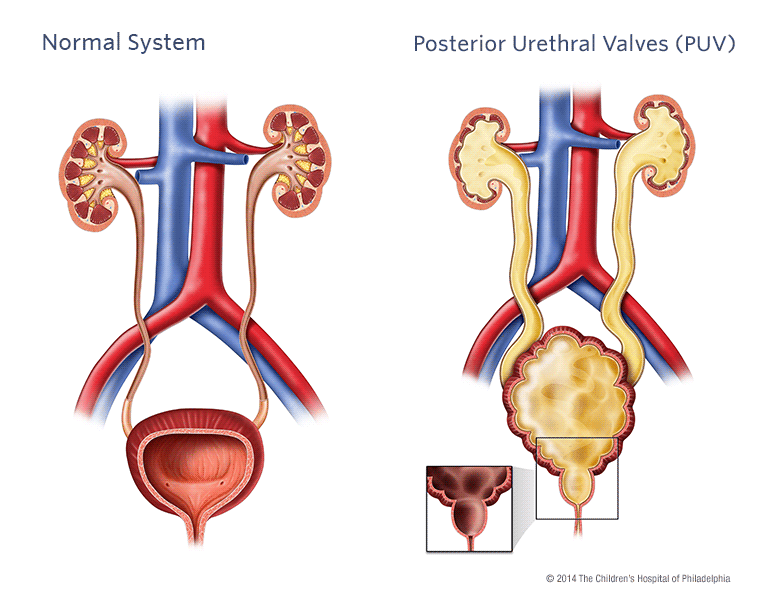
Posterior urethral valves (PUV) are obstructive membranes that develop in the urethra (tube that drains urine from the bladder), close to the bladder. The valve can obstruct or block the outflow of urine through the urethra. When this occurs, the bladder, ureters and kidneys become progressively dilated, which can lead to damage.
Causes of posterior urethral valves
PUV are thought to develop in the early stages of fetal development. The abnormality affects only male infants and occurs in about 1 in 8,000 births. This disorder is usually sporadic (occurs by chance). However, some cases have been seen in twins and siblings, suggesting a genetic component.
Signs and symptoms of PUV
PUV occur in varying degrees from mild to severe. Due to increased use of prenatal imaging, PUV may be identified before any symptoms are present. If any dilation (hydronephrosis) is identified, your baby will be monitored throughout the pregnancy and after birth.
Surgical treatment
- Valve ablation: Once PUV are identified, they need to be surgically incised. During valve ablation, the urologist will insert a cystoscope, a small device with a light and a camera lens at the end. He will use this instrument to make incisions in the valves so they collapse and no longer obstruct the urethra.
- Vesicostomy: In a situation where your baby is too small to undergo valve ablation or when a severe obstruction is noted, a vesicostomy may be recommended. A vesicostomy provides an opening to the bladder, so that urine drains freely from the lower abdominal opening.